

E-mail address: [email protected]
Popular choices
- Casinos Not On Gamstop
- Casinos Not On Gamstop
- UK Betting Sites
- UK Online Casinos Not On Gamstop
- UK Online Casinos Not On Gamstop
- Non Gamstop Casinos UK
- Slots Not On Gamstop
- Sites Not On Gamstop
- UK Casino Not On Gamstop
- Non Gamstop Casinos
- UK Online Casinos Not On Gamstop
- UK Online Casinos Not On Gamstop
- UK Online Casinos Not On Gamstop
- Casinos Not On Gamstop
- Top UK Slot Sites
- Casinos Not On Gamstop
- Non Gamstop Casino Sites UK
- Sites Not On Gamstop
- Casino Sites UK Not On Gamstop
- Casinos Not On Gamstop
- Casino Sites Not On Gamstop
- UK Casinos Not On Gamstop
doi:10.1016/S0531-5131(03)01095-1
Click here for the PDF version
Embryological development of the laterocervical region is closely linked with the branchial apparatus. Branchial arches are arranged in pairs on both sides of the midline. Morphologically, this apparatus can be described as a succession of folds and grooves corresponding to arches and clefts. On the other hand, a cleft refers to the ectoblastic furrow, and a pouch to an entoblastic one. An arch corresponds to a whole fold. Each arch gives rise to a bony and/or cartilaginous derivative, one or more striated muscles, a mixed cranial nerve and a vascular component. During development, the second arch expands downward to meet and merge with the fifth covering third and fourth ones to form the cervical sinus. On an anatomical point of view, a tract with two open-ends is called a "fistula", a dead-end tract is called a sinus, and lesion contacting neither ectoblastic nor entoblastic plate is a cyst.
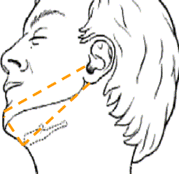
First branchial arch anomalies are quite uncommon lesions. One of the most important series reports 39 cases [1]. Clinically, Work proposed a classification, which is both anatomical and histological. In this classification, there are two types of malformations: type 1 presents as a cystic mass and are strictly ectodermal while type 2 presents as a cyst, sinus or fistula (or any combination) and can be ectodermal as well as entodermal or mixed. In our opinion, we think that things are often more complex. For example, authors such as Belenky and Medina [2] or Aronsohn et al. [3] are unable to classify more than 40% of their cases, and Triglia et al. 37% [1]. Our experience suggests that analysis of clinical manifestation and the findings of careful physical examination paying special attention to the external auditory canal are more helpful than anatomical or histological classification for management of these malformations. Clinically, first branchial arch anomalies are usually located on a triangular area between external auditory canal, the tip of the chin and the hyoid bone (Fig. 1). Any tumor or fistula in this area must suggest a first branchial arch anomaly. Two other typical clinical manifestations are, first a membranous attachment between the floor of the external auditory canal (EAC) and the tympanic membrane, and second a fistula on the floor of the cartilaginous part of EAC. The membranous attachment (Fig. 2) is asymptomatic and found in 44% of cases [1], but the fistula (Fig. 3) can present as an otorrhea. The main radiological investigation is CT imaging which allows the physician to appreciate the relationship between tumor and external auditory canal, and the region of the facial nerve trunk. In Fig. 4, correlation between CT scan and a cystic first arch anomaly is shown. Treatment is based on surgery. Because of the vicinity of the facial nerve, it has to be performed through a parotidectomy approach. The tract can be medial or lateral to the facial nerve, or swinging between its branches. The other important point of this surgery is the external auditory canal. Special care has to be taken in order not to miss any part of the tract. For that reason, the piece of cartilage through which the tract passes has to be removed. All authors have found a high incidence of recurrence in cases of previous inappropriate treatment: better said "the less child has been operated on, the better it is".
Second cleft anomalies are the most frequent branchial malformations. In Nicollas et al.'s series [4], 37 out of 68 branchial malformations were second cleft anomalies. These malformations are due to the persistence of the cervical sinus, and that explains why they are so close to the large vessels. Two clinical presentations can occur. The first and the main is a fistulous tract which ends in the skin of the anterior part of the SCM muscle, typically at the middle third. The malformation can be uni- or bilateral, and in that case the physician has to look for a branchio-oto-renal syndrome ordering an audiogram and a renal ultrasound. No radiological examination is necessary for this presentation. Treatment is surgical removal using two incisions (Fig. 5). The lower includes the cutaneous hole of the fistula and the upper incision is usually made at the level of the hyoid bone in order to help the dissection. Through this second incision the tract can easily be dissected over the carotid bifurcation, as far as possible. Resection can be bilateral if necessary without any risk or problem.
The second clinical presentation is an isolated cyst. Anatomically, it can present as a well-delineated lesion or include a tract-like extension on one side. Bailey has classified their anatomical extension in the neck into four types. Type I represent cysts under the superficial cervical fascia. On Type II, cysts are located in front of and lateral to the large vessels as shown in Fig. 6a. Type III cysts occur between the branches of the carotid artery, and type IV, as shown in Fig. 6b, are located between the large vessels and the pharyngeal wall. Surgical resection is performed through a latero-cervical approach and dissecting step by step. Recurrence is very uncommon.
Fourth branchial pouch anomalies theoretically correspond to the persistence of the pharyngo-branchialis duct. Why theoretically? Because we believe that it is more appropriate to talk about malformations of the "pyriform sinus area" than to differentiate third and fourth pouch anomalies. Third pouch fistulas end in the upper part of the pyriform sinus and go down, crossing over the superior laryngeal nerve while fourth branchial pouch anomalies fistula end at the apex of the pyriform sinus and go to the thyroid area crossing under the superior laryngeal nerve [5]. Sometimes, the fistula crosses over the nerve when it should have crossed under and vice versa. Clinically, those malformations account for about 1% of congenital cervical malformations [6]. Three clinical manifestations can occur. First is a latero-cervical abscess; usually, it is recurrent and left-sided. Some right-sided cases have been reported, but in more than 90% malformations are on left side. Second is pseudo-thyroiditis. There is no isolated infectious thyroiditis in children. The third aspect is a cutaneous fistula, which never occurs spontaneously but appears after an abscess. After looking at the neck and before obtaining a CT scan, endoscopy will often give the key of the diagnostic hypothesis. In most cases, the end of the fistula, which sometimes is infected, can be seen in the left pyriform sinus as shown in Fig. 7. The CT scan is the main radiological examination in this malformation. Fig. 8 shows a typical aspect of fourth pouch anomaly in contact with the left thyroid lobe. Surgical removal is performed through an anterior left-sided approach. The cyst and fistula will be dissected from the skin to the pyriform sinus mucosa. The fistulous tract can run behind or through the inferior cornu of thyroid cartilage. In this case, we resect the entire tract with the involved segment of cartilage. Embryologically, it corresponds to the tract passing through the chondrification area of the thyroid cartilage wing. If there is concern about clear identification of the recurrent laryngeal nerve, we think that it is necessary to perform a left thyroid lobectomy to visualize the nerve directly.
The embryologic origin of thymic cysts is unclear and several hypotheses are proposed. The two main theories are persistence of the pharyngothymic duct, or deterioration of Hassal's corpuscules [4, 7]. Clinically, Mikal proposed a classification in three types, which seems to be quite interesting: type 1 are "genuine" cysts and will be further described. Type 2 are thymic remnants with fistulas from skin to pharynx. Both types result in an abnormal persistence of the pharyngothymic duct. Type 3 cysts are fistulas originating from branchial apparatus and containing ectopic thymic tissue. Clinical aspect of "authentic thymic cysts" is a non-infected lateral tumor of the neck, more often left-sided than right-sided. CT scan is important because it identifies a retro-clavicular component of the tumor. After surgical removal, there is usually no recurrence.
In this review of congenital lateral cysts and fistulas of the neck, embryology, clinical aspects of the malformations, CT scan and surgery had been described. First and foremost ENT surgeons are physicians and "key symptoms" (Table 1) may help to direct the correct treatment to the correct patient. This is the key for avoiding recurrence in congenital malformations.
| Symptoms | Suggested anomaly |
|---|---|
| Otorrhea and/or tympanic membrane attachment | First arch malformation |
| Mass or fistula in parotid area | |
| Fistula on anterior border of SCM muscle | Second cleft malformation |
| Recurrent laterocervical abscess | Fourth pouch malformation |
| Thyroiditis | |
| Non-infected laterocervical ovoid mass | Thymic cyst malformation |