

E-mail address: [email protected]
Popular choices
- Casinos Not On Gamstop
- Casinos Not On Gamstop
- UK Betting Sites
- UK Online Casinos Not On Gamstop
- UK Online Casinos Not On Gamstop
- Non Gamstop Casinos UK
- Slots Not On Gamstop
- Sites Not On Gamstop
- UK Casino Not On Gamstop
- Non Gamstop Casinos
- UK Online Casinos Not On Gamstop
- UK Online Casinos Not On Gamstop
- UK Online Casinos Not On Gamstop
- Casinos Not On Gamstop
- Top UK Slot Sites
- Casinos Not On Gamstop
- Non Gamstop Casino Sites UK
- Sites Not On Gamstop
- Casino Sites UK Not On Gamstop
- Casinos Not On Gamstop
- Casino Sites Not On Gamstop
- UK Casinos Not On Gamstop
doi:10.1016/S0531-5131(03)01110-5
Click here for the PDF version
Contents
Diseased children are not frequently depicted in visual arts as in this drawing by George Grosz who, in 1935, sketched his little son Martin suffering from mumps (Fig. 1). Depictions of diseased and handicapped children were very rare before 1400, at approximately the beginning of the Age of Renaissance in Europe, but are increasingly found in the 19th century where children in general were portrayed more often. Diseased children illustrated in medical literature were not encountered before the 15th century.
It is often difficult to interpret a finding which deviates from our concept of normality as shown in the following relief (Fig. 2) from the New Kingdom in Ancient Egypt; an unusually intimate depiction of a king's family some 3345 years ago. King Akhenaten (or Aakunaten), on the left, is swaying his eldest daughter Merit-Amen with his hands and he is kissing her lips. His wife Nefertiti is holding her second daughter on her knees, while the youngest daughter is playing with her mother's pendants of the crown. All their daughters exhibit the same deformity of the head, similar to that of their father's head. It has not been clarified if this shape of the head is a symptom of a craniofacial dysostosis or the effect of bandaging of the infants' heads, as it was performed in other parts of the world, or a fashion of the art of the time.
The model head of the eldest daughter Merit-Amen (Fig. 3) shows the deformed head best. According to an ancient source King Akhenaten ordered his chief sculptor, Bek, to portray him in a way different from the standard representation. Thus, other interpretations concerning the King's distorted head seem to be somewhat speculative.
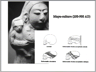
Fig. 4 (inset) shows several types of artificial deformation of the skull in infancy, a finding that can be observed in ancient cultures. For instance, the earthenware statuette of mother and infant from the Mayan culture (Fig. 4) gives us an idea how the modeling of the growing skull was achieved by means of a frontal board and bandages.
Albrecht Dürer was the first artist who systematically measured, as an epidemiologist would, about 300 persons including children, examining among other things the measures and proportions of a child as in the drawing of 1528 (Fig. 5).
Dürer also illustrated some deviations from the norm, as in his drawing "Mary with Jesus", (Fig. 6) where the diagnosis of rickets is very probable because of Jesus' square-shaped skull and the enlarged epiphyseal swelling of the arms and legs. Therapeutic options were poor at Dürer's time and subsequently, and were mostly confined to bloodletting, clysters, application of cups and cautery.
In the oil painting of Garemijn (Fig. 7), we can see how a physician practices clystering on an infant who probably also suffers from rickets (note his head and the epiphyseal areas of the limbs).
Until today, magical and fantastical ideas about positive and damaging influences have dominated the imagination of pregnant women concerning their unborn babies. The Swabian artist of this paper-maché (Fig. 8) gives us an idea of his "3-D Magnetic Resonance Image" of the foeti around 1440.
Therefore, one had to wait until the baby was born and, as this father of healthy twins exhibits, everybody was happy when they heard the newborn crying loudly (Fig. 9).
In the Middle Ages, a test of viability was to demonstrate the newborn's ability to cry so loudly that it could be heard in the four corners of a room. This was handed down in the illuminated manuscript of the "Sachsenspiegel", an illustrated book of law of Saxonia. Here, it reads: "Born alive is any child, if one can hear its voice at the four walls of the house". The half figures in the corners (Fig. 10) represent the statement that "one can hear its voice at the four walls of the house". The mother emphasizes this by pointing with her index finger. This is also one early depiction of the ability to hear by holding the ear forward with the index finger into the direction of the sound.
At one time, a common method of disposing unwanted and handicapped children was drowning, as shown in this illustration from Dijon (Fig. 11). Here, three women of various classes have the same goal: to hurl their unwanted children off a bridge into the swirling torrent below. The child of the woman to the right is almost drowned; only its legs and right arm are still rising from the water. The woman with the simple, blue dress is in the act of dropping her infant, weighed down with a stone, from the bridge while the richly dressed lady to the left is just stepping onto the bridge with her child in arm, in order to dispose of it "cleanly" in the river.
Inflammations of the upper airways, primarily diphtheria, were important causes of death in small children for hundreds of years. This toddler with perioral pallor fights for air and the outstretched hands of its mother vainly beg for help (Fig. 12).
Saint Blaise, an ancient physician in Sivas in Asian Turkey, is known as a guardian of the neck: "Blaise's blessings". He performed several miracles including saving a child who had choked on a fish bone. Before his beheading, Blaise beseeched God that anyone with a disease of the throat should be healed if he prayed to the saint with the words: "Blaise, martyr and servant of Christ, said 'either go up or go down'". This recommendation goes back to the Greek physician Aetius (5th century AD) who left the text in Latin: "Blasius martyr et servus Christi dixit: aut ascende, aut descende". Saint Blaise is surrounded by putti (Fig. 13) that display his attributes: the crozier, candle, and book. In front of the bishop there lies a putto who is being menaced by a huge toad crouching on his neck. This is certainly a very original allegory of a disease entity of the neck.
Since antiquity, ex-votos have been testimonies of faith in the form of bodily organs or pictures which were offered both as a plea for help and as thanksgiving. In this votive image (Fig. 14), the resuscitation of a girl is depicted who has fallen into a washtub. With the help of a little tube, a physician aspirates water from the mouth and pharynx. This salvation of the little girl seems to be ascribed to the Mother of God, who is depicted in a cloud.
The intubation of a diphtheric infant, 100 years later, is performed by a physician without the help of a saint as shown in this oil painting by the French artist Chicotot (Fig. 15).
The injury of the newborn's nose during the act of birth and its repair was described by Laurence Sterne in his book: "The Life and Opinions of Tristam Shandy, Gentleman" (Fig. 16). We read: "This unfortunate drawbridge of yours, quoth my father... God bless your honour, cried Trim, 'tis a bridge for master's nose. In bringing him into the world with his vile instruments, he has crushed his nose, Susannah says, as flat as a pancake to his face, and he is making a false bridge with a piece of cotton and a thin piece of whalebone out of Susannah's stays, to raise it up". We can see that the method of nasal reposition in newborns has remained the same, though with different instruments.
William Hogarth illustrated this scene in the book of Sterne (Fig. 17).
Otto Dix portrayed his son Ursus after his birth with a bluish-red swollen nose as a sequelae of a perinatal injury (Fig. 18).
In the Musée de l'Homme in Paris, there is a vessel from the Peruvian Moche culture depicting a mother with her baby (Fig. 19). The mother's midface shows destroyed nasal structures and a defect of the upper lip, while the nose and the lip of the baby appear normal, this supports the interpretation that the mother's midfacial defects, probably a sequelae of Leishmaniasis, were acquired after birth and not transmitted to the foetus.
William Hogarth tried to combat the spreading scourges of syphilis and alcoholism in England by depicting their catastrophic sequelae. He used three hallmarks in his art to indicate syphilis: saddle nose, other nasal defects, and black round patches applied to the skin to mask syphilitic sores. Let us have a look at Hogarth's last print (Fig. 20) of the series 'Marriage A-la-Mode', termed 'The Suicide of the Countess' with the detail of the countess's little daughter. The child is embracing her dead mother. Leg irons reveal that the child suffers from syphilis or rickets, while the black spot on the child's cheek shows that the fathers' sins have been visited upon the children. The child's high brow and saddle nose have also been interpreted as evidence of congenital syphilis.
Some congenital disorders are inescapable, and often signify a lifelong handicap, such as dwarfism. The cretin, Francisco Lezcano, was about 14 years old when Velásques portrayed him (Fig. 21). The mentally retarded dwarf runs his hands absently over the edges of the playing cards. The ENT doctor recognizes the facial hypoplasia on the right, a hypoplastic nose with stenosis of the nasal vestibule, and the open mouth.
Artists have depicted dwarfism, as in achondroplasia, for thousands of years. Fig. 22 shows an achondroplastic dwarf from the Late Period of Ancient Egypt with the typical frontal bossing, saddle nose, and short, deformed extremities.
In 1920, Otto Dix depicted the same disease in his painting "Praha Street" (Fig. 23).
In the painting "St. Bernard heals the sick" (Fig. 24), we see one of the diseased children holding a little bell in his right hand with his thumb and index finger. The other fingers seem syndactylic. The child's head is remarkable for its brachycephaly with frontal bossing, exorbitism and hypertelorism, the flat bridge of the short nose, and its deep root. These anomalies of the head, together with the malformation of the right hand, would suggest that the child suffered from Apert's syndrome.
Representations of Down syndrome are also known from early cultures, especially of Central America. An early illustration of this disease is displayed in the Museum of Aix-la-Chapelle, Germany in a child whose hair is being disordered by a monkey (Fig. 25). The child holds the animal on a neck chain. Its slumping shoulders and round face with characteristic facial dysmorphia are very similar to the symptoms of a child with Down syndrome. Apparently, the handicapped child and his monkey have been set out in the marketplace for the amusement of passersby.
In this watercolor, the Russian painter Alexandrowitsch Wssjewoloshskij caricatured a mother and her baby at the court of the tsar (Fig. 26) when he depicted the inherited progenia and enlarged lower lip, which were well-known characteristics of the Habsburgian dynasty.
One impressive natural history of the alterations of inherited progenic features during growth can be seen in the numerous depictions of the Emperor, Charles V, born 1500 in Ghent. Hans Holbein the Younger painted Charles at the age of 7 with a still straight nasal dorsum and a mild enlargement of the lower lip and chin (Fig. 27).
The oil painting of Charles V at age 16, by Bernard van Orley, shows an increased nasal hump, a severe progenia with angle class III malocclusion, and the protruding Habsburgian lower lip (Fig. 28).
An unknown artist painted Emperor Charles V at the age of 45 with marked Habsburgian characteristics, partly camouflaged by the beard (Fig. 29).
Patients suffering from a cleft lip and palate have been the subjects of artistic renditions since antiquity in many cultures. Almost true to life is the depiction of a bilateral cleft lip and palate found in a clay figure from the Central American Gulf Coast culture (Fig. 30, left). Through the wide-open beak of a bird, the anterior part of a human head is to be seen, exposing eyes painted with pitch. The short, slightly curved nasal dorsum ends in the tip, which leads down directly into the prolabium and protuberant premaxilla. The flattened and displaced left nasal wing opens the view to a left-sided cleft lip as part of an underlying bilateral cleft lip and palate. All the anatomical details of the cleft region and the nose are so painstakingly modeled that even a modern photograph would hardly be more precise as in the photograph of one of my patients (Fig. 30, right).
Among the works of the Russian painter Vrubel, I found the portrait of a sad boy with left cleft lip and nasal stenosis sitting in a baby carriage (Fig. 31). This is a very personal statement of a father and painter, whose child was born with an incomplete cleft lip and deformed left nasal wing.
In 1495 in Worms, Germany, two girls were born connected to each other in the frontal area, a so-called Metopagus (Fig. 32). One of the girls died at the age of nine. The physician then had to cut her off the still living twin, who later died due to the open frontal wound, as can be read in a contemporary report.
A rare malformation is the so-called Diprosopus, another stage of the incomplete separation of identical twins. This anencephalic cadaver specimen (Fig. 33, left) from the Institute of Pathology in Munich, which we studied 7 years ago, has got three eyes, two noses, two mouths, a double tongue base, and two auricles. This Mesoamerican clay figurine (Fig. 33, right) of a high valley of Mexico, dated about 900 BC, also presents with three eyes, two noses, two mouths, and two auricles. It is possible that the Mesoamerican artist had observed or heard of a diprosopic child to make this clay figurine approx. 2900 years ago.
Let us now look at some paintings where probably snoring and obstructive sleep apnea are depicted. Sleepiness is very apparent in the aspect of the overweight child depicted as a detail by Jordaens' in his large painting "Diogenes with a Lantern in Search of a Man at the Market" (Fig. 34). The child with its widely opened mouth and the large tongue also appears to be mentally retarded. It is possible that the painter had once seen a child with Prader-Willi syndrome, a syndrome that may be associated with sleep apnea.
At the same time, Juan Carreno de Miranda painted this portrait of the 5-year-old Eugenia Martenez who also looks as if she suffered from Prader-Willi syndrome (Fig. 35).
One of the pioneers of the 19th century who documented sleep-disturbing snoring in children was George Catlin, lawyer, civil engineer and noted painter of the American Indian. This drawing of a snoring boy (Fig. 36) is part of the booklet "Shut Your Mouth and Save Your Life". The comment to this drawing reads: "Children, I have said, are not born hunchbacks, but a habit of sleeping thus, in the varying temperatures of the night, might make them such. Infants are not born idiots or lunatics, but a habit of sleeping thus, in sudden change of weather, would tend to make them so".
A massive adenotonsillar hyperplasia, untreated in childhood, may result in a so-called long-face syndrome as can be seen in the features of this adolescent depicted by Jan Provoost (Fig. 37).
In the last century, the removal of the obstructing adenoids had become very popular like this unpleasant method to examine a child's nasopharynx by the physician (Fig. 38). Here the aggressive examination had been photographed for a German ENT book. The physician is probing the child's mouth, thrusting his index finger deeply into the throat. In order that the child cannot bite the palpating finger, the examiner's other hand pushes the skin of the child's cheeks between the rows of teeth.
This brutal method of examination is seen through the eyes of the Austrian artist Helnwein in his painting, "The Operation" (Fig. 39). A girl, her left hand tied down, lies supine on an operating table, fully clothed. A tight leather belt about her thorax and arms has committed her to immobility. The gigantic steel tube, which coming down from above has filled the child's mouth, underscores this state of being tied down. The girl is forced, defenceless and totally awake, to endure this torture. In the background, we see a similar scene as in the photograph of the ENT textbook.
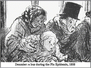
Let us now look at some other nasal diseases in art. It was a veritable passion of the French caricaturist Honoré Daumier to again and again treat the subjects of sniffles, flu and hankies in relation to curiously shaped noses in his lithographs. His 1858 lithograph "A Bus During the Flu Epidemic" not only shows suffering adults, but also a small child coughing his lungs out (Fig. 40).
In the 1920s, Heinrich Zille, a popular caricaturist in Berlin, depicted the most effective way to remove the snot from one's sister's nose (Fig. 41).
Wilhelm Busch has drawn an allergic response of the external nose to a bee sting in his comic strip series "The small thieves of honey" (Fig. 42). Peter and John wanted to satisfy their sweet tooth by stealing honey, but the bees stung into their noses, which resulted in an allergic edema.
The father tried to soothe them with cold water (Fig. 43). In vain, the boys could not eat their beloved dumplings, thus, the father had to call for the blacksmith of the village who removed the stingers with a huge pliers.
The physician put a black plaster on top of their noses (Fig. 44), and a happy ending came after a deep sleep when Peter and John could again enjoy their dumplings.
Artists also depicted the sequelae of an early nasal injury. Rembrandt, for instance, portrayed "The girl at a window" who has got a crooked hypoplastic nose to the left (Fig. 45, left) and Otto Dix painted the "Boy from the Working Class" with a bony deviation of the nasal pyramid, an asymmetric face, squinted eyes, a prominent left ear and a hopeless facial expression as symbols of the lad's social status during the hunger years following World War I (Fig. 45, right).
To this day, there is still controversy over when to operate on these fixed nasal deformities acquired during midfacial growth. The painting by Helnwein, entitled "Mean Child", depicts a terrified child who has just undergone a reconstructive operation to form a new nose from a frontal flap (Fig. 46). Blood drips from the tubes projecting from the reconstructed nose. A purulent scrap of granulation is seen in the left medial canthus, while a fresh scar from which the sutures have just been removed stretches from the angle of the mouth to the left ear. The flowered wallpaper in the background contains these words: disobedience allowed, taking pleasure in punishments, unchaste things, and other words which are connected with lines to the pathological alterations in the face. Do these harken back to the mediaeval belief that sickness is a punishment for greater or lesser human failings?
From the beginning of the 16th century, the use of a padded cap to prevent injuries of the head in infants during their period of learning to walk is documented in medicine and arts. In 1577, the physician Omnabonus Ferrarius recommended the head pad in his book on Pediatrics (Fig. 47): "... because it often happens to the child to fall down while beginning to walk. Therefore, to prevent injuries to the face and head, I recommend to put upon the child's head a round ring each day, made from wrapped linen or from buck's skin and filled with wadding, divided into four parts like a king's crown..." An outstanding example of prophylactic medicine more than 400 years ago!
The drawing of Rembrandt of an infant with head pad between two women (Fig. 48) and Rubens' oil painting depicting his son Peter Paul protected by a black head pad (Fig. 49) are also beautiful documents of prophylactic medicine from more than 300 years ago.
Epistaxis has often been depicted in visual arts. Therefore, I am presenting only a few examples. The wax figures of the artist Gaetano Guilio Zumbo which describe the horrors and sequelae of the plague, and which are on display in the Museo della Specola, Florence, seem to be taken from life, as in for instance the tiny stream of final nose bleed in a dead baby (Fig. 50).
In this ex-voto (Fig. 51), Saint Notburga with her typical symbols (sheaf, sickle, bunch of keys, bucket for milk) is asked for help with a child with nosebleed. The girl is sitting in an upright position on a stool, the head slightly bent forward so that the blood from her nose cannot spoil her clothes.
Heinrich Zille from Berlin, mentioned above, described a boy with a nosebleed who is weeping because he has injured his nose in a soccer game and returns to his mother in need of consolation (Fig. 52).
In the last 20 years, it has been the desire of many ambitious parents that their children should become famous sport stars, not considering possible long-term consequences. The Austrian artist Manfred Deix has provided us with an almost macabre cartoon of a baby in nappies being subjected to senseless training in boxing by his father who, in trying to ready him for the Olympic Games, has just given him a bloody nose (Fig. 53). The father's sarcastic consolation for his crying and bleeding son: "Stop crying 'Daddy, Daddy'?!?! For now I'm not your daddy, but your enemy... so beat me, stupid!!!"
Patron saints can be helpful – in many ways. Their multifacetted assistance is shown, for example, in the "Blessed Agostino Novello Altarpiece", of Simone Martini in Siena (Fig. 54). The altarpiece is composed of a central area with Agostino and of two side areas with Agostino's miracles. I would like to focus on two miracles: a child falling out of his bed (right side, bottom) and a child being attacked by a wolf when it was playing outside the gates of the city (left side, top). The scene of the falling child shows a very early depiction of epistaxis of an infant, dated 1324 AD. We look into the bedroom of a house where a pendulous bed is hanging, the torn suspension rope having made the infant fall out. A woman in a pink dress is calling for help by turning her eyes to Agostino, who is floating down through the air from the upper right corner.
The infant's head is embedded in a puddle of blood (Fig. 55). Its eyes are closed, and small separate streams of blood come from the closed mouth and from the nostrils. These nature-like details allow the diagnosis of an unconscious infant, the condition being caused by a severe skull injury with bleeds from the mouth and nose.
In the other miracle, a boy has been attacked by a wolf (Fig. 56). The child is lying on the ground and is bleeding from a bite wound to the left zygomatic and infraorbital region. The left eye has been evulsed from its socket. Alongside the child, there is the wolf flashing its teeth.
Very nature-like is this small polychrome semi-relief of Phoenician ivory from the 8th century BC in the British Museum (Fig. 57), part of a furniture ornament from Calah (Nimrud). In this semi-relief, a lioness has thrown an Ethiopian boy to the ground and is ripping open his throat.
The horrific abuse and slaughter of children by warriors is certainly as old as mankind itself. The archetype in Christian iconography is seen in "The Slaughter of the Innocents at Bethlehem", though the event is not at all historical. From the fifth century on, there are many depictions of how soldiers stab the children who also die due to neck injuries. In this example from Baroque, created by the leading mannerist among Dutch painters, Cornelis Cornelisz van Haarlem, the accuracy of the artist's eye is visible in this detail (Fig. 58) from his painting "The Murder of the Innocents". Alongside the deadly blade, fine drops of blood spray in a narrow gush from the infant's short neck in whose face there is nothing but pain and fear.
An illustration in the manuscript "Book of the Deeds" from the 15th century (Fig. 59) provides details of an inflammatory neck tumor suffered by a child of Rouen in 1281. The inscription tells that the tumor, which started the size of a chicken's egg, had ulcerated, became purulent and expanded as far as the larynx, then to the contralateral side of the neck and even into the armpits. The large lesion became clear of infection, but could not be healed.
In 1731, more than 20,000 Protestants were driven from the Archdiocese of Salzburg on account of their confession and were resettled in The Netherlands, Germany, and even in Georgia in the United States. As in many Alpine areas, goiter was endemic in the region of Salzburg. An example is shown with this engraving of the emigrant Steinbacher with her three children, of whom at least one girl has as much sign of a goiter as her mother (Fig. 60).
The "battered-child syndrome" is not an invention of our time. As early as the Middle Ages, we find documents telling of insane or drunken parents who injured their children with blows or even killed them. In 18th century England, many children were injured by their drunken parents. It was again William Hogarth who, as an artist, tried to fight against alcohol as here in the engraving "Gin Lane" (Fig. 61). Between 1730 and 1749, approximately 75% of all baptised children under 5 years died, very often due to alcohol embryopathy and early abuse of gin. Probably this microcephalic infant, thrown down to death by her drunken mother, had this symptom of alcohol embryopathy.
In this detail of the right edge of the engraving (Fig. 62), we see how a mother is feeding her infant with gin, and how two young orphan girls are drinking gin for breakfast, because they were too poor to get normal food.
The vertiginous effect of alcohol upon a child can be seen in this detail of a sarcophagus from the 3rd century AD where a staggering drunken boy, with a bunch of grapes in his hand, is supported by another boy (Fig. 63).
Vertigo can also be a symptom of a diseased ear, our last topic. Seven women carved in stone are part of the wall decoration in the entrance hall of the cathedral in Freiburg and represent the ideal of Christian education. One of them (Fig. 64, left) keeps a bundle of rods in her right hand ready to give the standing child the stick because he has not learnt his lesson unlike the sitting boy who is reading a book. She punishes the boy by pulling his left auricle, a method that I found as early as in the 4th century BC in a Greek terracotta from Tanagra. In 1841, Honoré Daumier caricaturized this instrument of education in his lithograph "How to Finally Get a Young Man to be Respectful to his Parents" (Fig. 64, right).
Marc Chagall has also depicted this pulling at children's ears in an etching (Fig. 65, left) from the series illustrating Gogol's "Dead Souls" (1924–1926) as commentary to the following text: ...and then there is that similarly well-known, but nonetheless unpleasant feeling when, after these words, his earlobes were painfully twisted by his father's fingers with their long nails. "Also, comics like to depict this method of education as we can see from the naughty boy Jeremiah in Lucky Luke's series "California or Death" (Fig. 65, right).
The removal of ear wax from a child's ear is rarely depicted in art as here in a woodcut of the Japanese painter Utamaro (Fig. 66).
Ferdinand Bol, a pupil of Rembrandt, depicted an example of a prominent ear (Fig. 67, left). The boy is also suffering from favus. Otto Dix painted another example of prominent ears in his work "Matchbox-seller" nearly 400 years later (Fig. 67, right).
Several persons with various grades of microtia are probably depicted in the altarpiece of the "Master of the Wenemaer Triptych" in the Museum for Fine Arts at Ghent. The artist painted all the people's faces with life-like features. Deformed auricles are visible in 9 out of a total of 41 persons of all age groups, among the auricles one microtic ear, which belongs to Jesus as a newborn, which is really unusual (Fig. 68).
In another scene of this altarpiece, we see Jesus with a normal left auricle and two angels with left-hand microtia (Fig. 69).
Fig. 70 probably shows two shepherds as examples for adults with microtia in this altarpiece. In the shepherd with a flute, the ear remnant is positioned extremely low. The neck is short and broad, the mandible seems underdeveloped, and there is some degree of hypertelorism. The bearded shepherd is painted with a left ear that is positioned low on the face. The pinna and the earlobe are underdeveloped, and adjacent to the deformed lobe, there seems to be a skin tag.
Dürer left a remarkable pencil drawing that depicts moderate microtia (Fig. 71). The upper half of the pinna of the child's right ear is severely deformed.
Coming to the modalities of treating hearing loss in children, we encounter, of course, more miracles than causative methods. The ancients ascribed the inner tempests of severely hearing impaired children who were also believed to be mute as being the work of demons, by which some mutes were possessed. This woodcut depicts St. Aldedrut (Fig. 72) helping a mute girl fettered to a column, a meaningful symbol of the girl's handicap. As the saint, reading from the Bible, is pointing with her fingers at the mouth of the girl, the demons of disease depart from her mouth, where they inhibited her speech.
This miniature to the initial 'O' (Fig. 73) of the artist Liberale da Verona shows Christ bending over the standing child, touching the child's right ear and lips with his hands in order that he might again hear and speak.
In this ex-voto dating to 1720 (Fig. 74), the cure of deafness is represented by two bright rays, which link Saint Mary (cut off in this detail) in the clouds with the ears of the child who lost his hearing after a severe illness, and received help. This ex-voto shows the mother in the foreground who also tries to help her son.
In the next ex-voto (Fig. 75), a mute boy is standing all alone in the countryside looking up to Saint Mary in the clouds for help. Beneath Mary, we read: "The child is mute, 1816".
To facilitate the ability of speaking in infancy, the frenulum was cut, a recommendation since the time of the Roman writer Celsus. The town physician of Ulm, Scultetus, described in his surgical textbook "Armamentarium Chirurgicum" which instruments were used to cut the frenulum off the tongue, namely a small scalpel and a special tongue forceps (Fig. 76). Many had to pay for this operation with their death due to bleeding or infection.
In Fig. 77, Jules Lenepveu depicts how Jacob Rodrigues Pereire, the first teacher for deaf-mutes in France, tries to teach the girl Marbois from Orléans to recognize tones via the vibrations of the larynx.
A miniature, with four events in the "Book of the Deeds of our Lord Saint Louis", tells the story of a boy in the 13th century who was deaf and mute by birth. The miracle happened at the grave of St. Louis. We see how the deaf boy is approaching the grave together with other diseased people (Fig. 78, left). On the right part of the figure, the boy is kneeling in front of the grave praying.
The miracle made the boy able to hear the bells (Fig. 79) and even more miraculously, he realized that he was able to speak, as noted by his lifted arms and his open mouth (Fig. 80).
In this presentation, you have seen a lot of miracles that happened due to prayer and the help of saints. In the last 100 years, more and more miracles in handicapped children have been achieved on the base of scientific progress, technical development, and surgical skill. What is the future? Will we still need miracles of the older type at all when all the details of diagnostics and treatments have been explained on a rational base one day? I think so, because healing which we physicians have experienced every day with our patients will not be possible without the help of an omnipotent creator.