bDivision of Pediatric Cardiothoracic Surgery, Cincinnati Children's Hospital Medical Center, USA
E-mail address: [email protected]
Popular choices
- Casinos Not On Gamstop
- Casinos Not On Gamstop
- UK Betting Sites
- UK Online Casinos Not On Gamstop
- UK Online Casinos Not On Gamstop
- Non Gamstop Casinos UK
- Slots Not On Gamstop
- Sites Not On Gamstop
- UK Casino Not On Gamstop
- Non Gamstop Casinos
- UK Online Casinos Not On Gamstop
- UK Online Casinos Not On Gamstop
- UK Online Casinos Not On Gamstop
- Casinos Not On Gamstop
- Top UK Slot Sites
- Casinos Not On Gamstop
- Non Gamstop Casino Sites UK
- Sites Not On Gamstop
- Casino Sites UK Not On Gamstop
- Casinos Not On Gamstop
- Casino Sites Not On Gamstop
- UK Casinos Not On Gamstop
doi:10.1016/S0531-5131(03)01072-0
Click here for the PDF version
Contents
1. Commentary
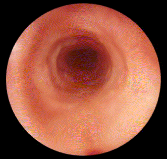
Complete tracheal rings are rare, and historically are associated with a high mortality. There are several methods for managing children with complete tracheal rings, and our unit has extensive experience with most of these. We commenced utilizing the tracheal slide technique for operative repair of complete tracheal rings in February 2001, and the same surgical team operated on 10 children with complete tracheal rings over the next 18 months. Some of these children were extremely tenuous, with multiple congenital anomalies. Nine were repaired under cardiac bypass. In all cases, the tracheal surgery was successful. There was one late death not related to the tracheal repair.
We have found slide tracheoplasty to be vastly superior to previous methods of tracheoplasty that we have utilized for the management of complete tracheal rings (Figs. 1–18).